What Makes Wisdom Teeth Removal More Complex?
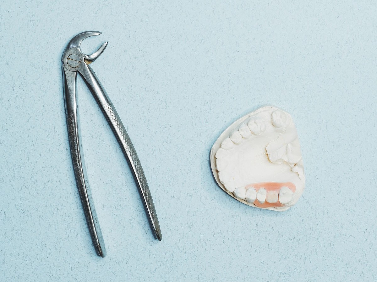
Wisdom teeth removal sounds straightforward until you look at the details that actually determine how simple or difficult the procedure will be. Two patients can both be told they need their wisdom teeth out, yet one may have a quick office-based extraction with mild soreness for a few days, while the other needs a longer surgical procedure, a more involved recovery, and careful follow-up. The difference usually comes down to anatomy, timing, and how the teeth relate to the surrounding bone, gum tissue, nerves, and sinuses.
In practice, complexity is rarely about a single factor. It is usually the accumulation of several small issues that turn a routine case into a technical one. A partially erupted lower wisdom tooth in a healthy 17-year-old is not the same problem as a fully impacted tooth in a 38-year-old whose roots wrap close to the inferior alveolar nerve. Both fall under the broad label of Wisdom Teeth Removal, but they are different procedures in almost every way that matters.
Patients often think complexity must mean danger. That is not necessarily true. A complex removal can still be predictable and safe when it is properly planned. What complexity changes is the level of surgical judgment required, the amount of imaging and preparation needed, the type of anesthesia that may be appropriate, and the aftercare that follows.
It starts with position, not pain
One of the most common misunderstandings is that painful wisdom teeth must be the hardest to remove. Pain does not tell the whole story. Some of the most difficult extractions involve teeth that have never caused obvious symptoms. Meanwhile, a tooth that hurts because of gum irritation may be relatively easy to remove because most of the crown is already visible in the mouth.
The exact position of the tooth matters more than the amount of discomfort. Oral surgeons look at whether the tooth is fully erupted, partially erupted, or fully impacted. They also consider the angle at which it is growing. A wisdom tooth can lean forward into the second molar, tilt backward, lie nearly sideways, or remain buried upright under bone. Each pattern changes how access is gained and how much bone or tooth structure needs to be removed.
Lower wisdom teeth tend to be more technically demanding than upper ones. The bone in the lower jaw is denser, which often means the tooth is held more firmly. Visibility can also be more limited in the back of the lower jaw, especially in patients who have smaller mouths, strong cheek muscles, or limited ability to open widely. A surgeon may need to section the tooth into pieces rather than trying to remove it whole. That is not a sign that something has gone wrong. It is often the safest and most controlled way to proceed.
Bone changes everything
When a wisdom tooth is covered by bone, the procedure shifts from a simple extraction to a surgical one. That distinction matters. Soft tissue can be reflected and repositioned with relative ease, but bone requires deliberate removal and carries more postoperative consequences. More bone removal usually means more swelling, more soreness, and sometimes a slightly longer recovery.
The density of the bone matters too. Younger patients often have somewhat more forgiving bone and roots that are not fully developed. In contrast, older patients usually have denser bone and more fully formed roots, sometimes with curves or divergent tips that make the tooth resist removal. This is one reason many surgeons prefer to evaluate wisdom teeth in the late teens or early twenties. It is not because every patient must have them removed early, but because surgery can become more complicated as the surrounding structures mature.
I have seen this difference play out clearly in practice settings where younger siblings come in after an older brother or sister delayed treatment for years. The younger patient may have four impacted teeth removed in a single visit and recover with typical swelling and a few quiet days. The older sibling, with denser bone and roots set deeper around critical anatomy, often needs a more cautious procedure and reports a slower return to normal chewing and jaw function.
Root shape is a hidden variable
The crown is what patients imagine when they think about a tooth, but surgeons worry just as much about the roots. Roots are not always neat, straight, or separate in a simple pattern. Wisdom teeth are notorious for variation. Some roots are fused into a conical shape, which may actually simplify removal. Others curve sharply, flare apart, or hook around bone in ways that increase resistance.
This matters because the path the tooth must travel during removal is limited. If a tooth has roots that spread like the legs of a stool, it may not come out in one piece without removing more bone than is ideal. In those cases, sectioning the tooth is a strategic choice. The crown can be divided from the roots, and the roots can sometimes be removed individually with less stress on the jaw and surrounding tissues.
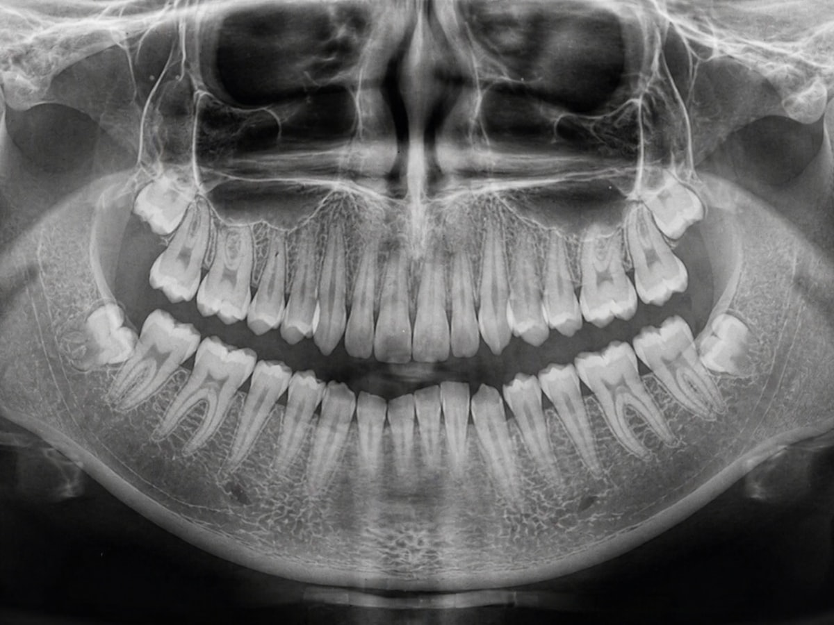
Radiographs help identify these patterns, but even good imaging has limits. A tooth can look manageable on a two-dimensional panoramic image and reveal unexpected root curvature once surgery begins. That is one reason experienced surgeons talk about planning in terms of probabilities rather than guarantees. Good judgment comes from expecting variation and having options ready when the anatomy turns out to be less cooperative than the image suggested.
Nerve proximity raises the stakes
For lower wisdom teeth, one of the most important questions is how close the roots sit to the inferior alveolar nerve, which runs inside the mandible, and the lingual nerve, which lies in the nearby soft tissue and provides sensation to part of the tongue. The closer a wisdom tooth is to these nerves, the more complex the procedure becomes.
Complexity here does not mean a nerve injury is likely. It means the surgeon must think carefully about the safest approach. Sometimes a panoramic radiograph shows warning signs, such as darkening of the root, diversion of the nerve canal, or loss of the canal outline. When those features appear, a cone beam CT scan may be recommended. Three-dimensional imaging can show whether the nerve runs to the side of the roots, below them, or in some cases in direct contact.
That information can change the plan. A surgeon might modify how the tooth is sectioned, how much force is used, or whether a coronectomy is worth discussing. In a coronectomy, the crown is removed and the roots are intentionally left behind when the roots are judged to be at high risk of damaging the nerve if fully extracted. It is not appropriate in every case, but it illustrates how complexity sometimes requires a more nuanced solution than simply taking the whole tooth out.
Patients are often surprised to hear that less can sometimes be more. Surgical success is not measured by aggressiveness. It is measured by solving Wisdom Teeth Removal the problem while preserving function and minimizing avoidable risk.
Upper wisdom teeth have their own challenges
Upper wisdom teeth are often easier to remove, but they are not automatically simple. Their main anatomical relationship is with the maxillary sinus. If the roots are very close to the sinus floor, or if part of the tooth projects toward the sinus, extraction requires careful technique to avoid creating a communication between the mouth and the sinus.
Most small sinus exposures heal well when managed properly, but the possibility changes the conversation before surgery and the instructions after it. Patients may be advised to avoid forceful nose blowing or anything that builds pressure for a period after the extraction. If sinus involvement is likely, the surgeon plans the flap design, instrument choice, and closure with that in mind.
Upper wisdom teeth can also be awkward because their position in the far back of the mouth limits visibility. A tooth that is high, tilted, or partly hidden behind the second molar can be hard to access without stressing the adjacent tooth or soft tissue. Complexity is not always dramatic. Sometimes it is simply a matter of angle, access, and the discipline to work carefully in a confined space.
The age of the patient matters more than many realize
Age influences almost every dimension of Wisdom Teeth Removal. In late adolescence, the roots may be only partially formed, the bone is often less dense, and healing tends to be quicker. By the late twenties, thirties, and beyond, extractions can still be done very successfully, but they often demand more time, more surgical manipulation, and more recovery.
There are biological reasons for this. With age, bone becomes less elastic and roots become fully developed. Teeth may also become ankylosed in rare cases, meaning they are fused more directly to bone and do not move as expected. The surrounding periodontal condition can change as well. Gum recession, adjacent restorations, existing fillings on the second molar, and prior episodes of infection all add complexity that simply is not as common in younger patients.
This is one reason delayed removal can be a mixed decision. Waiting is sometimes reasonable, especially when wisdom teeth are healthy, functional, and easy to monitor. But if there is already evidence of recurrent inflammation, decay, or damage to the neighboring second molar, delay can convert a manageable operation into a more involved one. The trade-off is individual. Good care depends on matching the timing to the anatomy and the patient's broader oral health, not following a one-size-fits-all rule.
Infection and inflammation can make surgery harder
Active infection changes tissue behavior. Inflamed gum tissue bleeds more easily, local anesthesia may be less effective in highly acidic infected areas, and opening the mouth may be uncomfortable if the patient has trismus, or limited jaw motion, from inflammation. Even before the surgeon begins, visibility and access may already be compromised.
Pericoronitis is a classic example. A partially erupted lower wisdom tooth develops a flap of gum over the crown, food and bacteria collect under it, and the area becomes swollen and tender. Some cases calm down with irrigation, hygiene measures, and time. Others flare repeatedly. When surgery is done during or after repeated episodes, the tissue can be more fibrotic, more vascular, and more difficult to handle cleanly.
Chronic low-grade infection can also affect the tooth next door. It is not uncommon to see decay begin on the back side of the second molar where a poorly positioned wisdom tooth traps plaque in a space that is nearly impossible to clean. Once the neighboring tooth is involved, the situation is no longer just about removing a problematic third molar. The surgeon and general dentist may need to coordinate restorative care as well, and the consequences of delay become more significant.
Access is a real surgical issue
People tend to think of tooth extraction as a matter of grip and force. In reality, access is often the deciding factor. A patient with a small oral opening, pronounced gag reflex, thick cheeks, a large tongue, or jaw joint stiffness can turn an otherwise ordinary case into a much more technically demanding one.
Limited opening narrows the angle available for instruments and makes it harder to visualize the back of the mouth. This is particularly relevant for lower wisdom teeth, where the surgeon needs enough room to reflect tissue, remove bone, section the tooth if needed, and retrieve fragments without pushing them into unfavorable positions. If the patient is very tense, fatigue and muscle resistance can further reduce access during a long procedure.
Sedation can help in selected cases, not just for anxiety but for muscle relaxation and procedural control. That said, sedation is not a magic fix. It improves conditions, but it does not change the underlying anatomy. It is one tool among many, and the decision to use it should be based on the expected difficulty of the case, the patient's medical history, and how safely the procedure can be performed in the planned setting.
Medical history can complicate a dental problem
Wisdom teeth exist in a dental space, but surgery happens in a medical one. A patient's health status can make an extraction more complex even when the tooth anatomy is favorable. Blood thinners, immune suppression, poorly controlled diabetes, history of radiation to the jaws, bisphosphonate therapy, and certain cardiac conditions all shape surgical planning.
Sometimes the complication is logistical rather than technical. The extraction itself may be simple, but coordinating with a physician, adjusting medications, or choosing the right setting for anesthesia adds layers of planning. For a healthy young adult, the procedure may be done comfortably in an office with local anesthetic and perhaps intravenous sedation. For a patient with significant systemic disease, the same extraction might call for a different environment or more conservative staging.
Here are a few medical factors that commonly influence complexity:
- Bleeding risk from anticoagulants or clotting disorders
- Delayed healing related to diabetes, smoking, or immune compromise
- Medication-related bone concerns, especially antiresorptive drugs
- Airway or anesthesia considerations that affect sedation choice
- Existing infection that may spread more readily in vulnerable patients
None of these automatically prevents surgery. They simply mean the plan has to be tighter, the communication clearer, and the margin for improvisation smaller.
Imaging often determines whether a case is routine or not
A basic panoramic x-ray remains an excellent screening tool, but some wisdom teeth demand more detail. When the relationship to the nerve is uncertain, the sinus anatomy looks unusual, or the root pattern is difficult to interpret, three-dimensional imaging can shift the entire risk assessment.
This is one of the places where patients occasionally mistake thoroughness for upselling. In reality, additional imaging is usually recommended because the surgeon sees a specific anatomical question that two-dimensional films cannot answer well enough. Knowing whether a root is merely close to the nerve or actually superimposed over it can influence consent, technique, and the discussion of alternatives.
Good imaging does not make surgery easy, but it makes surprises less likely. That matters because most complications in oral surgery do not come from lack of technical skill alone. They come from anatomy behaving differently than expected. Better information before the procedure often leads to gentler decisions during it.
Recovery is more complex when the surgery is more involved
Patients judge difficulty partly by what Wisdom Teeth Removal happens after the appointment, and for good reason. More extensive surgery usually means more swelling, more jaw stiffness, and a slightly higher chance of postoperative issues such as dry socket, prolonged soreness, or food trapping in the healing site.
Lower impacted wisdom teeth, especially those requiring bone removal and tooth sectioning, tend to produce the most memorable recoveries. Swelling often peaks around the second or third day, not immediately. Chewing may feel awkward for several days. Some patients notice temporary bruising along the jawline or under the chin. These are not necessarily signs of trouble. They are often the normal consequences of a controlled surgical procedure in dense tissue.
The harder part is distinguishing normal recovery from a problem. Persistent worsening pain after an initial improvement, foul taste, fever, marked asymmetry, or numbness that does not begin to fade on the expected timeline deserve reassessment. Clear postoperative instructions matter because they help patients identify what is expected and what is not.
What surgeons look for before calling a case complex
Complexity is usually recognized before the first instrument is picked up. A thorough evaluation often reveals the likely pressure points. Surgeons are typically thinking about several questions at once:
- Is the tooth buried in soft tissue, bone, or both?
- How close are the roots to the nerve or sinus?
- Will the tooth need to be sectioned for safe removal?
- Does the patient's age or medical history change healing or anesthesia planning?
- Is there enough access to do the procedure safely and efficiently?
That kind of preoperative thinking is what separates a routine visit from a carefully staged surgical event. From a patient's perspective, it may look like extra x-rays, a longer consultation, or a referral to an oral surgeon. From a surgical perspective, it is risk management in its proper form.
A difficult extraction is not a failed one
There is an unfortunate tendency to judge dental procedures by speed. If a wisdom tooth takes longer to remove, patients sometimes assume something unusual happened. More often, the opposite is true. A surgeon who slows down to section a tooth carefully, preserve bone where possible, and avoid excessive force is usually doing exactly what the anatomy requires.
The goal is not to prove how quickly a tooth can be removed. The goal is to remove it with the least trauma necessary. That may mean making a larger flap for visibility, trimming a small amount of bone to create a path of removal, or stopping short of full root removal when nerve risk outweighs the benefit. Those choices reflect judgment, not hesitation.
A straightforward case can turn tricky, and a seemingly intimidating x-ray can end in a smooth procedure. Experience helps, but so does humility. The best operators respect the anatomy, plan for variation, and communicate honestly about what the images do and do not reveal.
Why one person's story should not set your expectations
People compare wisdom teeth stories constantly. One friend says they went back to work the next day. Another says recovery lasted two weeks and the swelling was dramatic. Both accounts may be accurate, and neither tells you much about your own case unless the anatomy and health context were similar.
A 19-year-old with partially formed roots, soft tissue impaction, and no medical issues is starting from a very different place than a 34-year-old with fully impacted lower thirds close to the nerve and a history of repeated pericoronitis. Even the same patient can have very different experiences from side to side. One lower wisdom tooth may come out with modest effort, while the other requires more bone removal because of its angle or root form.
That variability is why meaningful consent includes specifics. Patients benefit most when they hear not just the generic risks of Wisdom Teeth Removal, but the reasons their own teeth fall into a lower-risk or higher-complexity category.
The real answer is anatomy plus judgment
What makes wisdom teeth removal more complex is rarely the label itself. It is the combination of impaction, root shape, nerve or sinus proximity, bone density, inflammation, access, age, and medical history. Add these factors together, and the procedure may shift from simple extraction to meticulous oral surgery.
That does not mean a patient should be alarmed. Complexity is not a verdict. It is a planning category. When the anatomy is understood, the imaging is appropriate, and the surgical approach matches the case, even difficult wisdom teeth can often be managed predictably and safely. The important thing is not whether the tooth is called impacted or surgical. The important thing is whether the person treating it has recognized the details that make it different, and built a plan around those details rather than around assumptions.
Oxnard Dentistry
1730 E Gonzales Rd, Oxnard, CA 93036, United States
+1 805-307-8731
FAQ About Wisdom Teeth Removal
How painful is it to remove a wisdom tooth?
Local anesthetic numbs the area during extraction. Pressure may still be noticeable; tell the dentist if you feel pain. Soreness and swelling afterward are common. Follow the pain-control instructions provided for your situation.
How long is recovery for wisdom teeth extractions?
Recovery varies with the tooth position and complexity of the extraction. Some people resume routine activities quickly, while others need several days of rest. Soreness, swelling and jaw stiffness can last up to two weeks. Follow your clinician's aftercare and activity advice.
What are the risks of wisdom tooth removal?
Possible complications include dry socket, infection, persistent bleeding and changes in sensation from nearby nerve injury. Your dentist or oral surgeon should explain your individual risks before treatment. Seek prompt dental advice for bleeding that will not stop, fever, or severe or worsening pain and swelling.